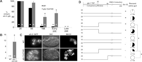
Figure 1.

Extended period of developmental plasticity in embryos lacking GLP-1/Notch function, and temporal requirement for GLP-1. (A) Proportion of terminal embryos with ectopic elt-2∷GFP in wild-type (WT) and glp-1(e2144) embryos following induction of END-3 at the indicated stage. “Early 20E” indicates that END-3 was induced in embryos shortly after the last E descendants were born (approximately “bean” to “comma” stage). “Late 20E” indicates that END-3 was induced at approximately the “twofold” stage of morphogenesis, based on time from the two- to four-cell stage. Numbers in bars indicate total embryos carrying the hs-end-3 array scored. (B) Proportion of terminal embryos with ectopic expression of the late gut differentiation marker IFB-2, detected with antibody MH33 (Zhu et al. 1998) following induction of END-3 at the 20E stage. (C) Expression of intestinal differentiation markers following late activation of END-3 in embryos of the indicated genotype. (D) Timelines of temperature shift experiments. Embryos were held at the indicated temperature [permissive (15°C) or nonpermissive (25°C) for the glp-1(e2144) mutation] and shifted at the indicated developmental stage. (Top) The known temperature-sensitive period for GLP-1 lethality (Priess et al. 1987) and the normal period of developmental plasticity (“competency window”) are shown on the developmental time line. lin-12(−) indicates that the gene was knocked down by RNAi in the mothers of the embryos where indicated. Pie diagrams show the proportion of embryos with ectopic endoderm; the total number of embryos carrying the END-3 transgene is indicated below each. In B–D, hs-end-3 was activated at a stage equivalent to the 20E stage in wild-type embryos. Error bars represent standard error. (*) Fisher's exact test, P < 0.01.
