Figure 2.
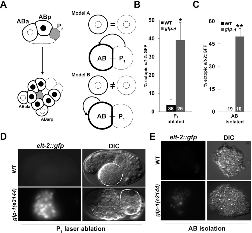
Notch signals regulating developmental plasticity are specific to the AB lineage. (A, left panel). Summary of early known cell fate-inducing Notch signals activated by P1 descendants. Gray-filled cells, derived from P1, signal their AB-derived Notch-expressing neighbors. Black outlines indicate cells expressing the GLP-1/Notch receptor. Cells in which Notch signal transduction has been received by signaling from P1 descendants contain black nuclei. (Right panel) Alternative models for Notch-dependent regulation of developmental plasticity. Circles at the top of each model represent isolated AB cells with (heavy lines) or without (light lines) GLP-1 present. Model A: Inductive signals from P1 descendants both activate cell fates and restrict developmental plasticity. Model B: AB-derived signals, distinct from the cell fate-inducing signals arising from P1 descendants, restrict developmental plasticity. (B) Proportion of P1-ablated embryos from wild-type and glp-1(e2144) embryos that express ectopic elt-2∷GFP following late induction of END-3 expression. (C) Proportion of partial embryos obtained from in vitro culturing of physically isolated AB blastomeres from wild-type and glp-1(e2144) embryos that express ectopic elt-2∷GFP. (D) elt-2∷GFP expression in wild-type and glp-1(e2144) embryos in which AB was isolated by laser ablation followed by late induction of END-3 expression. Ablated blastomeres are denoted by dashed lines. (E) elt-2∷GFP in partial embryos obtained from physically isolated AB blastomeres. In all experiments, END-3 was induced by heat shock when the number of cells corresponded approximately to the 20E stage in intact wild-type embryos. In B and C, numbers in the bars represent the total number of embryos carrying the END-3 transgene. (*) Fisher's exact test, P = 0.01; (**) Fisher's exact test, P < 0.01. Error bars represent standard error.

