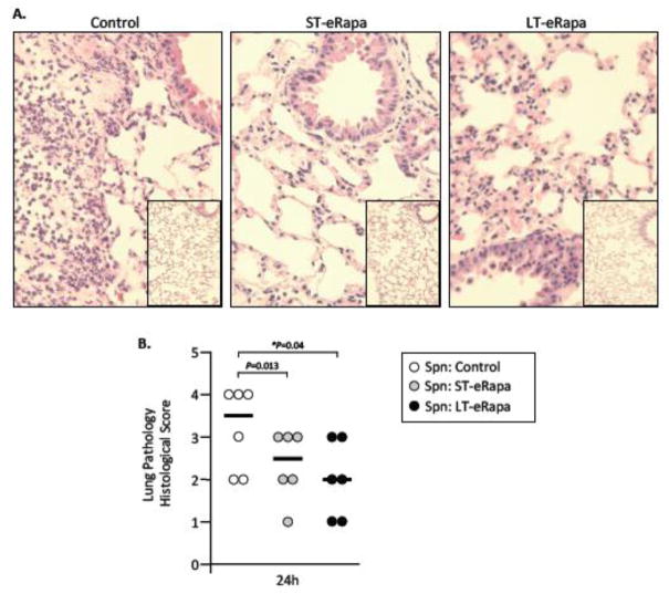
Figure 4. Impact of eRapa on lung pathology.
A) Representative micrographs of H&E stained lung sections from infected and mock-infected aged mice on control, ST- and LT-eRapa diets 24 hours post-intratracheal challenge with S. pneumoniae (n=6 per cohort; infectious dose 1.0 × 10⁴ cfu). Micrograph of mock-infected mice are inset within the larger image from infected mice. B) Lung histopathological scores for each infected mouse (individual circles) at 24 hours post-infection. Lungs were scored 0–5 on the basis of peribronchial and perivascular inflammation, neutrophil infiltration, and alveolar consolidation. For each mouse two separate non-adjacent lung sections were examined. The horizontal bar indicates the median score for the cohort. Statistical analysis was performed using one-way ANOVA comparing eRapa groups versus the control.

