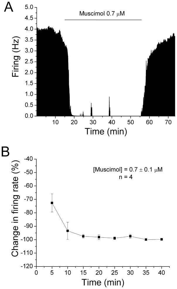
Figure 3. No reversal of muscimol-induced inhibition.
A. Mean ratemeter graph of the effects of muscimol (0.7 μM) on the firing rate of a single DA VTA neuron. Vertical bars indicate the firing rate over 5 sec intervals. Horizontal bars indicate the duration of muscimol application (concentrations indicated above bar). Muscimol initially produced a decrease in firing rate of 63.53% at 5 min, and the inhibition increase further so that by the end of the 40 min muscimol administration, the inhibition of firing rate was 99.96%.
B. The mean firing rate of 4 DA VTA neurons during the 40 min muscimol administration in experiments similar to the one depicted in Figure 3A. Muscimol concentration was adjusted so that greater than 50% inhibition was produced at 5 min; the mean concentration of muscimol that was administered was 0.7 ± 0.1 μM. Muscimol produced an inhibition in firing rate of 72.6% at 5 min and the inhibition increased significantly over time (one-way repeated measures ANOVA, F(7,21) = 7.16, p < 0.05).

