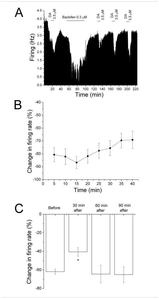
Figure 5. Desensitization to dopamine inhibition following reversal of baclofen inhibition.
A. Mean ratemeter graph of the effects of dopamine (3.5 μM) and baclofen (0.3 μM) on the firing rate of a single DA VTA neuron. Vertical bars indicate the firing rate over 5 sec intervals. Horizontal bars indicate the duration of dopamine or baclofen application (concentrations indicated above bar). Initially, a 5 min dopamine administration produced inhibition of 59%. During the 40 min application, baclofen produced a decrease in firing rate of 73% at 5 min, with a maximum inhibition of 84%, and inhibition of only 57% by the end of the 40 min period. During the 30 min washout period, the baseline stabilized at a new, lower level. Dopamine was administered for 5 min at 30 min intervals after the cessation of baclofen treatment, producing inhibitions of 29% at 30 min, 43% at 60 min, and 45% at 90 min.
B. The mean firing rate of DA VTA neurons during the 40 min baclofen administration in experiments similar to the one depicted in Figure 3A. Baclofen concentration was adjusted so that greater than 50% inhibition was produced; the mean concentration of baclofen that was administered was 0.29 ± 0.04 μM. There was a statistically significant reduction in baclofen-induced inhibition over time (One-way repeated measures ANOVA, F7,42 = 4.63, p < 0.05).
C. The effect of 5 min application of dopamine during and after the 40-min baclofen application period. The inhibitory effect of dopamine was significantly reduced only at 30 min after the 40 min baclofen administration period compared to the effect of dopamine before the baclofen treatment (One-way ANOVA, F3,29 = 12.01, p < 0.05).

