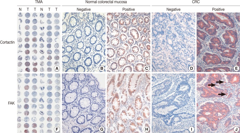
Fig. 1.
Immunohistochemical staining of cortactin (A-E) and focal adhesion kinase (FAK) (F-J) in microarray tissues of normal colorectal mucosa and colorectal adenocarcinoma (CRC). Tissue microarray (TMA) slide (A, F) is represented by one core of normal colorectal mucosa (N) and two cores of CRC (T). Most normal colorectal mucosas are negative for cortactin (A, B) and FAK (F, G). Some normal colorectal mucosas are positive for cortactin (C) and FAK (H), however, the intensity of cytoplasmic expression is weak to moderate. Some CRCs are negative for cortactin (A, D) and FAK (F, I), whereas most are positive for cortactin (E) and FAK (J) with more intense staining than those of normal colorectal mucosas. Note the expression of FAK on peritumoral blood vessels (J, arrows).

