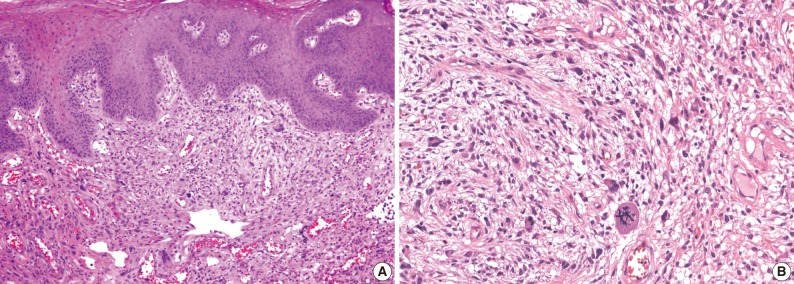
Fig. 1.
Microscopic findings of a cellular pseudosarcomatous fibroepithelial stromal polyp. (A) This fibroepithelial polyp of the vagina shows a single pedunculated mass covered with squamous epithelium. The stromal component shows variable cellularity from area to area. The subepithelial stroma is composed of atypical spindle cells with abundant blood vessels. There are also scattered stellate, bizarre multinucleated giant cells extending into the overlying mucosa. (B) The tumor cells of the pseudosarcomatous area show abundant pale cytoplasm with prominent nucleoli, coarse chromatin, nuclear pleomorphism, and numerous atypical mitoses.

