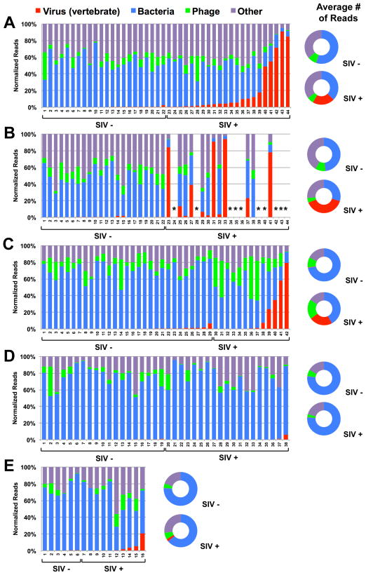
Figure 1. Taxonomic distribution of sequences identified in feces of SIV-infected (SIV+) and uninfected (SIV−) monkeys.
A–E graph the percentage of sequences obtained from fecal samples assigned by MEGAN to the indicated taxonomic groups. X-axis numbers refer to individual animals.
(A, B) Sequences from monkeys housed at NEPRC for 24 weeks (A) or 64 weeks (B) after intrarectal infection with SIVmac251. *euthanized for progressive AIDS 24 to 64 weeks after SIV infection.
(C) Sequences from monkeys housed at the TNPRC 23–64 weeks after intravaginal infection with SIVmac251.
(D) Sequences from SIV-infected and control vervet African green monkeys housed at NIH after intravenous infection with SIVagm90, SIVagmVer1 or after natural infection in the wild.
(E) Sequences from sabaeus African green monkeys housed at NEPRC and infected intravenously with SIVagmMJ8 or SIVagm9315BR.
(A–E) Flanking doughnut charts display the averaged values per kingdom for SIV+ or SIV- monkeys.
See also Figure S2

