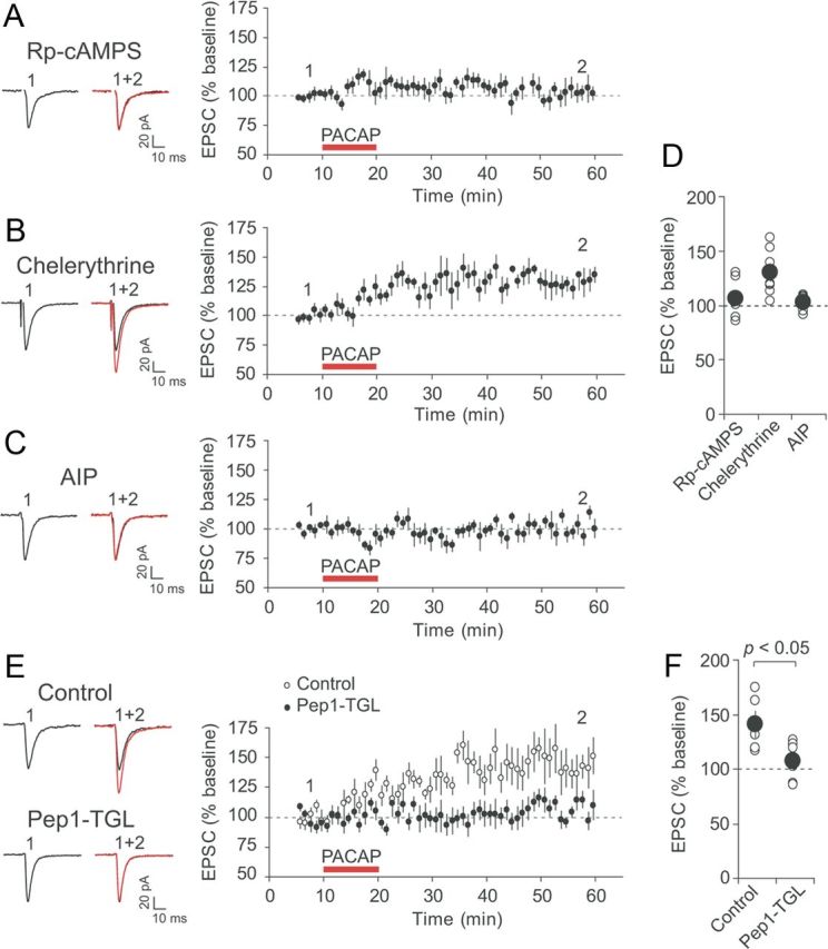
Figure 6.

Postsynaptic mechanism of PACAP38-induced synaptic potentiation in CeL neurons. A, Left, Examples of EPSCs recorded in CeL neurons before (black trace) and after PACAP38 application (red trace) when pipette solution contained the cAMP antagonist Rp-cAMPS (100 μm). Right, Time course of EPSC amplitude changes produced by PACAP38 (red horizontal bar) in CeL neurons loaded with Rp-cAMPS (n = 7). B, Effects of PACAP38 were tested in the presence of PKC inhibitor chelerythrine (10 μm in external solution) (n = 7). C, Effects of PACAP38 were tested in CeL neurons loaded with CaMKII inhibitor AIP (5 μm; n = 6). D, Summary of the EPSP amplitude changes after PACAP38 application for the experiments shown in A–C (n = 7 for the experiments with Rp-cAMPS; n = 7 for chelerythrine; n = 6 for AIP). E, Left, Examples of EPSCs recorded in CeL neurons before (black traces) and after PACAP38 application (red traces) under control conditions and when pipette solution contained Pep1-TGL (200 μm). Loading postsynaptic CeL neurons with Pep1-TGL blocked synaptic potentiation by PACAP38. This indicates that synaptic targeting of GluR1 subunit-containing AMPA receptors was necessary for PACAP-induced potentiation in CeL neurons. Right, Time course of EPSC amplitude changes produced by PACAP38 (red horizontal bar) in CeL neurons. F, Summary of the EPSP amplitude changes produced by PACAP38 application under control conditions and in the presence of Pep1-TGL in pipette solution (control, n = 5; Pep1-TGL, n = 9). Error bars indicate SEM.
