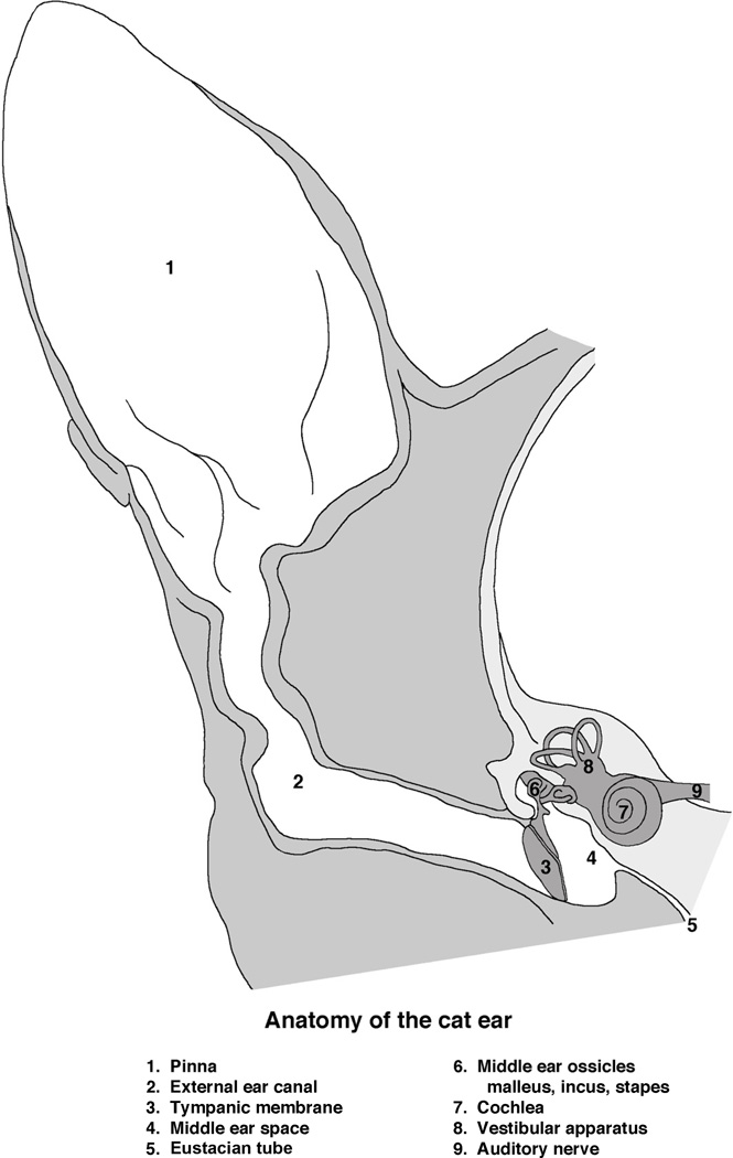
Figure 1.
Schematic drawing of the cat ear. The external ear consists of the pinna (1) and external ear canal (2) that conducts airborne sound to the tympanic membrane (3, ear drum). The tympanic membrane and three middle ear bones (6) occupy the middle ear space (4). These moving parts convert vibrations in air to vibrations in the inner ear (7). The middle ear space is confluent with the pharynx by way of the Eustachian tube (5). Behind the cochlea, the auditory component of the inner ear, lies the vestibular structures (8). The eighth cranial nerve, the auditory-vestibular nerve (9), conducts sensory information from the sense organ to the brain. (Drawn by Catherine Connelly, Garvan Institute of Medical Research, Sydney, Australia.)

