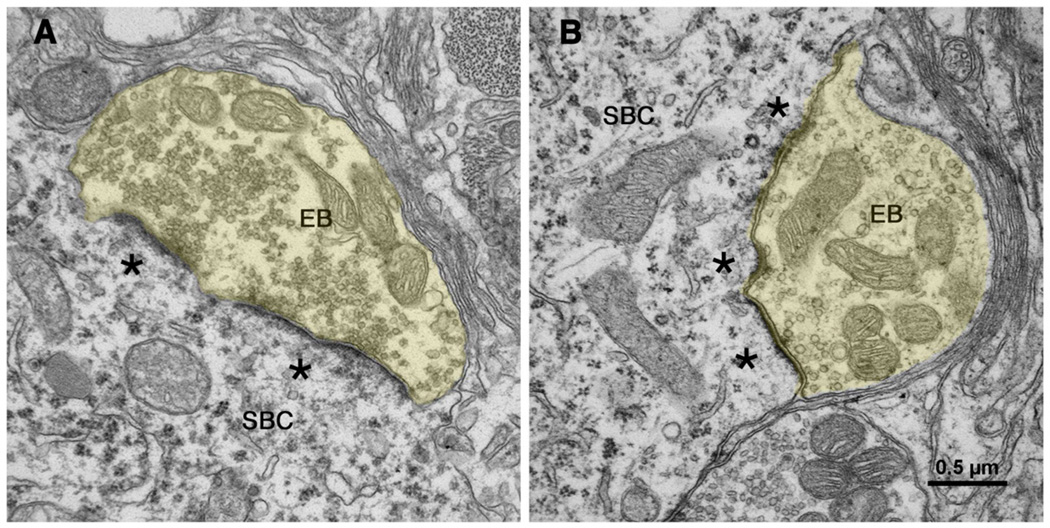
Figure 12.
Electron micrographs through synapses of endbulbs (EB) of congenitally deaf cats [135]. The postsynaptic densities of these synapses have hypertrophied (*) and become more flattened. Synaptic vesicles have proliferated in the endbulb (yellow) cytoplasm and intermembraneous channels have disappeared. The scale bar equals 0.5 µm. [135] (O'Neil, JN, Limb, CJ, Baker, CA, et al. Bilateral effects of unilateral cochlear implantation in congenitally deaf cats. J. Comp. Neurol. 2010;518: 2382–2404, with permission.)

