Figure 3.
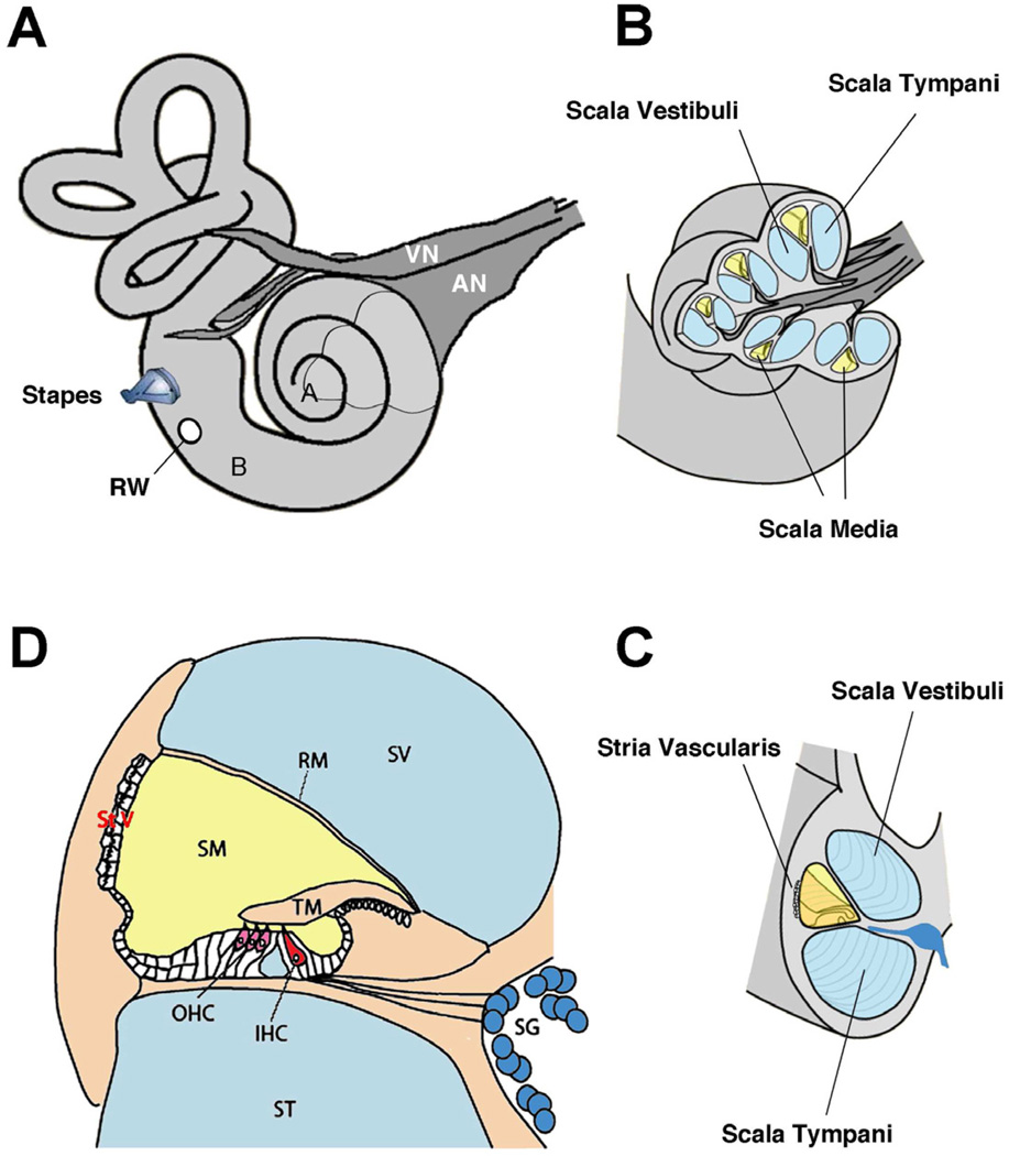
Anatomy of the inner ear. (A) view of right hearing and balance apparatus. The cochlea is the coiled structure on the right and the semicircular canals of the vestibular system are on the left. For the cochlea, “A” indicates the apex (low frequencies) and “B” indicates the base (high frequencies). The stapes, a middle ear bone, inserts into the vestibule of the inner ear; the round window (RW) is covered by a membrane that relieves the pressure when the stapes “pistons” into the ear. The auditory (AN) and vestibular (VN) nerves bundle together to form the 8th cranial nerve. (B) A section of the otic capsule has been cut away (indicated in A) to reveal the three chambers of the labyrinth. The sensory organ resides in the scala media (yellow). (C) A rotated view of the cut end of a cochlear turn showing the three chambers, with the scala media (yellow) and the stria vascularis. (D) Enlarged diagram showing a cross-section through the scala media, emphasizing the organ of Corti and the hair cell receptors.
Abbreviations: IHC, inner hair cell; OHC, outer hair cells; RM, Reissner’s membrane; SG, spiral ganglion; SM, scala media; ST, scala tympani; StV, stria vascularis; SV, scala vestibuli; TM, tectorial membrane. Adapted from Fig. 1, Eisen and Ryugo, 2007. (Eisen MD, Ryugo DK, Hearing molecules: contributions from genetic deafness, Cell Mol Life Sci 2007; Mar 64(5): 566-80, with permission.)

