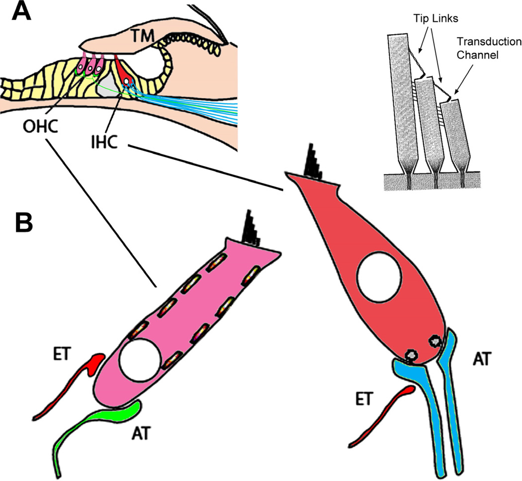
Figure 5.
Components of the organ of Corti. (A) The organ of Corti rests on the basilar membrane, and is composed of the sensory receptor cells (OHCs and IHCs), supporting cells (yellow) and the tectorial membrane (TM). (B) The hair cell receptors are innervated by afferent type I (blue) and type II (green) terminals as well as by efferent (ET, red) terminals whose cell bodies reside in the brain stem. At the apical ends of the receptor cells are stereocilia that form part of the transduction apparatus with tip-links and channels (upper right). Adapted from Fig. 1, Eisen and Ryugo, 2007. (Eisen MD, Ryugo DK, Hearing molecules: contributions from genetic deafness, Cell Mol Life Sci 2007; Mar 64(5): 566-80, with permission.)

