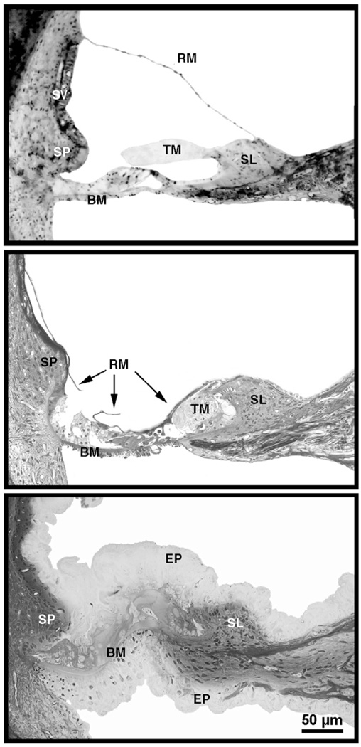
Figure 7.
Photomicrographs of organ of Corti in cats with normal hearing (top), cats deafened by the collapse of Reissner’s membrane, thinning of the stria vascularis, and obliteration of the sensory epithelium (middle), and cats deafened by spongioform hypertrophy that destroys the organ of Corti (bottom) [65]. Scale bar equals 50 µm. Abbreviations: BM, basilar membrane; EP, epithelium; RM, Reissner’s membrane; SL, spiral limbus; SP, spiral prominence; SV, stria vascularis; TM, tectorial membrane. (from Ryugo, DK, Cahill, HB, Rose, LS, et al. Separate forms of pathology in the cochlea of congenitally deaf white cats. Hear. Res. 2003;181: 73–84, with permission.)

