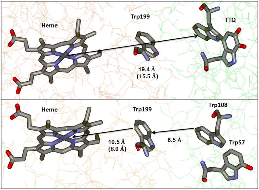
Figure 4.
Schematic representation of (Top panel) electron tunneling from the nearest heme of diferrous MauG to quinone MADH and (Bottom panel) hole hopping via Trp199 from preMADH to the nearest heme of bis-Fe(IV) MauG. Distances to and from the heme are listed both as to and from the heme Fe and the porphyrin ring (in parentheses). The figure was drawn using the coordinates from PDB ID: 3L4O (top) and 3L4M (bottom).

