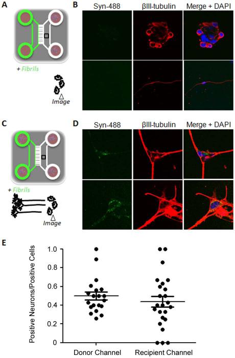
Figure 4.
Axonal transport and transmission of α-synuclein fibrils to second order neurons. (A) Set up of the control experiment to determine background retrograde transport. Syn-488 fibrils were added to one chamber that did not contain neurons. At the same time, recipient neurons were plated in the opposite channel that was given an additional 100μl of medium to prevent diffusion. (B) After four days of incubation the cells were stained with an anti-class III ß-tubulin (ßIII-tubulin) antibody to identify neurons and imaged by confocal microscopy. The upper and lower panels show two representative fields. (C) Schematic of the experimental setup to detect and measure transfer. Neurons were plated and cultured for one week to allow them to extend their axons to the opposite channel. At that time, Syn-488 fibrils were added to the chamber containing these neuron soma and freshly isolated neurons were added to the opposite channel. After one or four days in culture, depending on the experiment, the cells in both channels were stained with an anti-ßIII-tubulin antibody and analyzed by confocal microscopy. (D) The upper and lower panels show two representative fields. (E) For the experiments schematized in C, the percentages of Syn-488-positive cells that were neurons was determined by a blinded experimenter for the first neuron channel and the recipient channel. Graphs show individual values as well as mean ± SEM.

