
Fig 4.
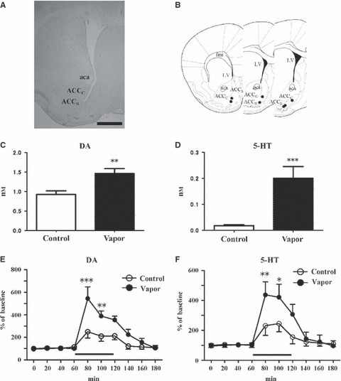
Releases of DA and 5-HT and effects of alcohol perfusion into the ACCS. (A) Placement of brain microdialysis guide cannulae or probes. All guide cannulae or probes were aimed at the right nucleus ACCS. Scale bar: 1 mm. (B) Black circles represent the guide cannula or probe tip as visualized by the manual inspection of Nissl-stained brain sections after collecting the dialysate from the ACC through the dialysis probe membrane. (C) Basal physical concentration of DA in the dialysate monitored by microdialysis in the ACCS after alcohol vapor exposure for 20 consecutive days (n = 6). **P < 0.01 compared with the alcohol-naïve control groups (n = 6; two-tailed unpaired t-test). (D) Basal concentration of 5-HT in the dialysate monitored by microdialysis in the ACCS after alcohol vapor exposure for 20 consecutive days (n = 5). Basal 5-HT release in the control group was at the limit of detection using our ECD-HPLC assay (n = 5). ***P < 0.005 comparison with the alcohol-naïve control groups (two-tailed unpaired t-test). (E, F) Time courses of changes in the extracellular levels of DA (E) and 5-HT (F) in the ACCS of the control (open circles; n = 5) and alcohol vapor-exposed mice (filled circles; n = 6) after the perfusion of 100 mm ethanol for 60 min. After establishing a stable baseline, 100 mm alcohol was perfused with the artificial CSF for 60 min through the microdialysis probe membrane (bar). *P < 0.05, **P < 0.01 and ***P < 0.005 (compared with the control mice; one-way anova and Bonferroni’s post hoc t-test). Values are means ± SEM. 5-HT, serotonin; aca, anterior commissure anterior part; ACCC, nucleus accumbens core; ACCS, nucleus accumbens shell; DA, dopamine; fmi, forceps minor corpus callosum; LV, lateral ventricle.