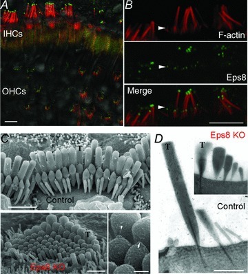
Figure 4. The actin-binding protein, Eps8, regulates hair bundle morphology.

A, IHC and OHC stereocilia showing immunostaining for F-actin (red) and Eps8 (green) from adult mice using confocal microscopy. Fluorescence image is superimposed on the differential interference contrast image. B, high-magnification image of IHC stereocilia showing that Eps8 is localized in the tips of stereocilia. Scale bars in A and B represent 5 μm. C, scanning electron micrograph showing the hair bundle structure in adult IHCs from control (top panel) and Eps8 knockout mice (Eps8 KO; bottom left panel). Taller stereocilia are indicated by T. Control hair bundles are normally composed of three rows of stereocilia. In Eps8 knockout mice, hair bundles are disorganized and shorter and additional rows of stereocilia are present compared with wild-type mice. Bottom right panel shows the presence of tip links between stereocilia (arrow and arrowhead; Eps8 knockout IHC), which are required for gating the mechanoelectrical transducer channels. Scale bars represent 2 μm (top panel), 1 μm (bottom left panel) and 250 nm (bottom right panel). D, transmission electron micrographs showing the stereociliary bundle from control and Eps8 knockout adult IHCs. Note that in this case the Eps8 KO IHC shows an extra row of stereocilia compared with the control cell. Scale bar represents 1 μm. Figure modified from Zampini et al. (2011).
