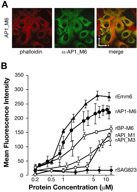
Fig 3.

Binding of rAP1_M6 and other recombinant proteins to epithelial cells. A. Confocal microscopy analysis of rAP1_M6 protein stained with specific antibodies (green) binding to epithelial cells (labelled with phalloidin, red); the x- and y-axis arrows in the merged image indicate 27 µm dimensions. B. FACS analysis of GAS proteins binding to A549 cells. Cells were incubated with increasing concentrations of the following recombinant proteins: pilus ancillary proteins rAP1_M6, rAP1_M1 and rAP1_M3, the pilus backbone rBP_M6, the rEmm6 protein and, as negative control, rSAG0823 from S. agalactiae. Cell-bound proteins were detected with specific antibodies and fluorescent secondary antisera, followed by flow cytometry analysis. Results are presented as the mean and standard deviation values of three independent experiments.
