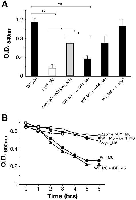
Fig 4.

Role of AP1_M6 in biofilm formation and bacterial aggregation. A. Crystal violet biofilm formation assay. GAS WT_M6, Δap1_M6 and complemented bacteria were grown for 10 h in 24-well plates, washed with PBS and stained with crystal violet. Experiments with WT_M6 were also conducted in the presence of purified mouse polyclonal antibodies raised against rAP1_M6 and rBP_M6 pilus components, and against rScpA as control. The mean and SD values of three independent experiments run in triplicate wells are reported. Significant differences between strains calculated by Student's t-test are shown by asterisks, **P < 0.01, *P < 0.05. B. Sedimentation of GAS WT_M6 grown in the presence or absence of 1 µM rAP1_M6 or rBP_M6, compared with that of Δap1_M6 mutant strain. Bacteria were grown for 16 h, bacterial precipitates were suspended by mixing, and OD values were at 600 nm measured at different time intervals under static conditions; the presented data derive from one of three experiments with reproducible results.
