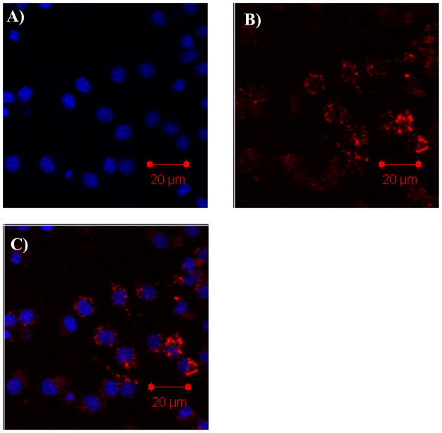
Figure 9.
The confocal images of RAW 264.7 cells incubated with R in micelle 2 (dye concentration is 4 μM and polymer concentration is 0.1 mg/mL) showing the particulate distribution and localization of the R after 16 h internalization. Blue emission represents the Hoechst 33342 (A). Red emission represents the R (B). The superimpose of the blue and red fluorescence is given in C.

