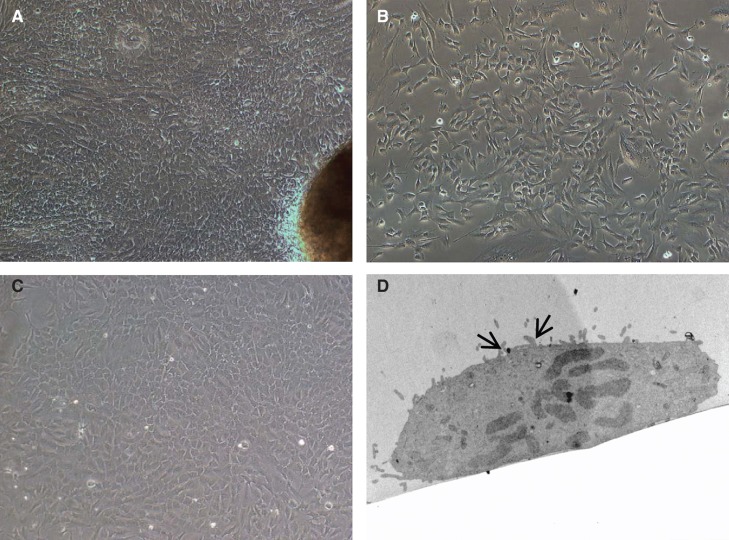
Figure 1.
Morphology of cells isolated from human small bowel. (A) Five to six days after placing tissue pieces in culture plates, cells started to migrate out from (B) a preconfluent culture of EpCAM-positive cells. These cells grew and showed epithelioid morphology and could be maintained for 6–7 passages. (C) Confluent monolayer of EpCAM-positive cells showing cobble-shaped morphology typical of epithelial cells. (D) Transmission electron micrographs of cultured primary enterocytes showing presence of microvilli (arrows). Magnification 20×.

