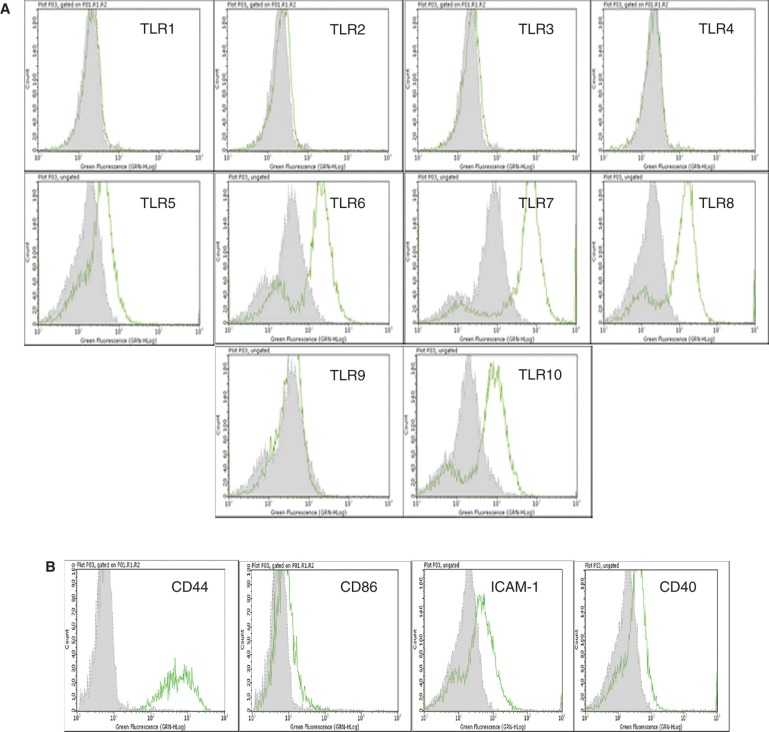
Figure 2.
Flow cytometric analysis of cultured human EpCAM-positive cells. (A) Flow cytometric results are presented in the form of histograms. Representative histograms for expression of TLRs in enterocytes are shown. Primary enterocytes did not express TLR-1, -2, -3, -4 and -9. They weakly expressed TLR-5, but demonstrated strong expression of TLR-6, -7, -8 and -10. The expression of TLRs was observed only in saponin-treated cells. (B) Isolated human enterocytes also stained positive for several immune recognition molecules such as CD44, CD86, ICAM-1 and CD40. Staining with only the secondary antibody served as negative control (grey filled histogram).

