
Figure 2.
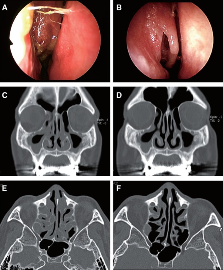
Endoscopic findings of right side nose and CT (coronal and axial) images in case no. 5. (A), (C), and (E) show status before steroid therapy. CT showed bilateral opaque areas located in maxillary and ethmoid sinus with a diffuse liquid pattern. Nasal discharge and nasal crusting were observed. (B), (D), and (F) show status after steroid therapy; these finding showed improvement of sinusitis, respectively.