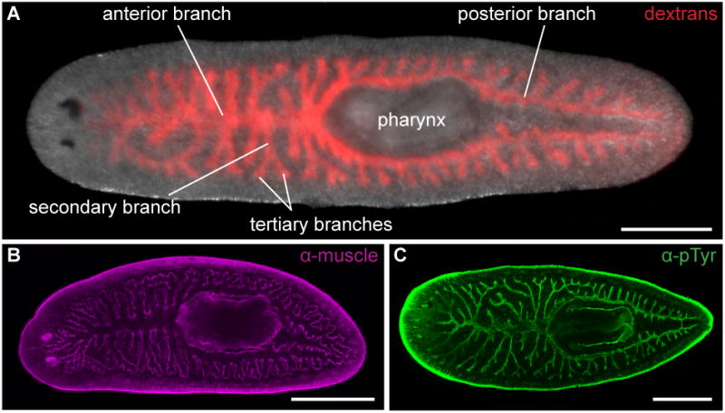
Figure 1. Anatomy of the planarian intestine.

(A) Animals were fed Alexa 546-conjugated dextrans (red) to visualize all intestinal branches. The intestine possesses three primary branches – one anterior and two posterior – from which secondary and tertiary branches project laterally. These branches meet at a junction just anterior to the pharynx, the muscular feeding organ through which food enters the intestine and waste is excreted. (B) Enteric muscles (magenta) surround all intestinal branches. The anti-muscle antibody also labels pharyngeal and outer body wall muscles. (C) Anti-phosphotyrosine (green) labels the lumenal surface (specifically apical cell-cell junctions) of intestinal epithelial cells. B and C are confocal projections. All panels are dorsal views, with anterior to the left. Scale bars, 500 μm.
