
Figure 6. Intestinal tissue remodels during regeneration.
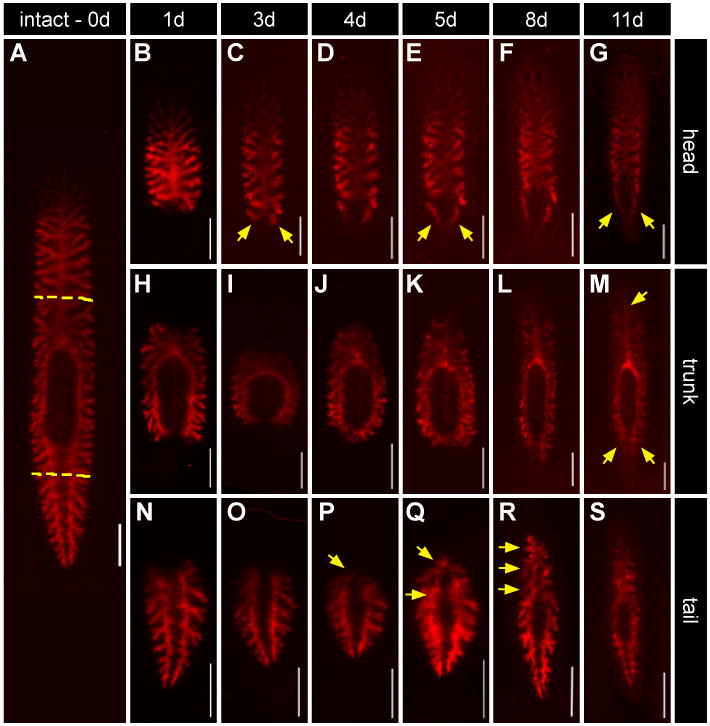
(A) A single animal was fed Alexa 546-conjugated dextrans (red) three times to label differentiated phagocytes. Animals were then amputated one day after the last feeding (yellow dashed lines). Living tissue fragments were imaged on subsequent days after cutting (top panel indicates times). (B-G) Head fragments. Labeled intestinal cells begin to project posteriorly as early as 3 days post amputation (C), contributing significantly to peripharyngeal and posterior branches by 11 days (yellow arrows, C, E, and G). (H-M) Trunk fragments. Anterior and posterior branches gradually elongate over the regeneration time course (yellow arrows, M). (N-S) Tail fragments. Labeled branches project across the midline initially at 4-5 days (yellow arrows, P and Q). Anterior branches eventually (R) coalesce at the midline to reconstitute the primary anterior branch. All scale bars = 500 μm.