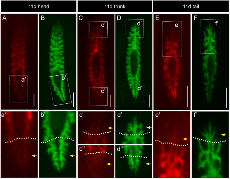
Figure 7. Differentiated intestinal tissue labeled prior to injury contributes minimally to regenerated branches within the blastema.

As in Figure 6, single animals were fed Alexa 546-conjugated dextrans (red, A, C, and E, insets in a', c'-c″, and e') to label differentiated phagocytes, and then amputated. At 10 days of regeneration, animals were fed Alexa 488-conjugated dextrans (green, B, D, and F, insets in b', d'-d″, and f') to visualize the entire regenerated intestine. All regenerates were imaged again at 11 days, one day after being fed green dextrans. (A-B) Head fragments. The most posterior regions of tail branches within the blastema (a', yellow arrows, compare to b') fluoresce only weakly in the red channel. (C-D) Pharyngeal fragments. Red fluorescence is reduced in both anterior and posterior branch tips (c'-c″, yellow arrows, compare to d'-d″). (E-F) Tail fragments. Red fluorescence intensity is lowest in the anterior-most branches (e', yellow arrow, compare to f'). Dashed yellow lines in a'-f' delineate the plane of amputation. All scale bars, 500 μm.
