Figure 3.
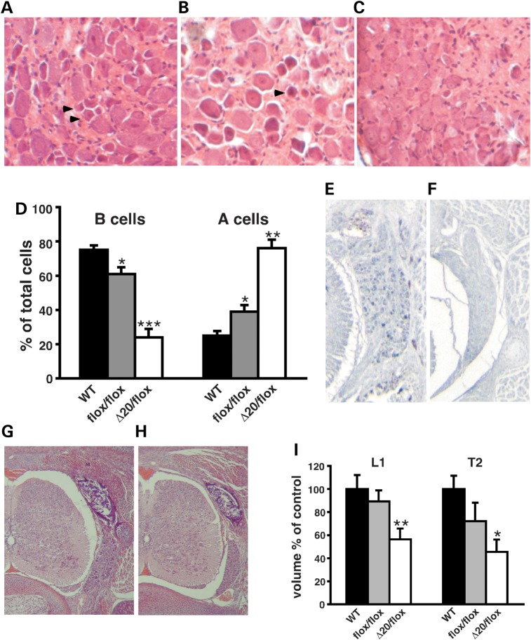
Sensory ganglia deficits in FD mouse models. (A–C) Representative H&E-stained cross sections through thoracic DRGs of 18-month-old WT (A), Ikbkapflox/flox (B) and IkbkapΔ20/flox (C) mice. Note that the DRGs of WT and Ikbkapflox/flox mice contain numerous small dark neurons (arrowheads), which are virtually absent in IkbkapΔ20/flox DRGs. (D) Quantification of small dark (type B) and large light (type A) neuronal profiles in 18-month-old WT, Ikbkapflox/flox and IkbkapΔ20/flox DRGs (n = 3). Percentages of type A and type B neuronal cells over the total number of cells are displayed (n = 3). (E and F) Transverse paraffin sections of E18.5 WT (E) and IkbkapΔ20/flox (F) embryos were immunostained with anti-CGRP antibody. Note the large number of CGRP-positive neuronal cells in WT DRG in contrast to their near absence in IkbkapΔ20/flox mutant DRG. (G and H) H&E-stained transverse sections of WT (G) and IkbkapΔ20/flox (H) E18.5 embryos show that the DRG is significantly reduced in the size in the mutants already at this stage. (I) Volumes of lumbar L1 DRG (L1) and thoracic T2 (T2) of E18.5 WT (n = 4), Ikbkapflox/flox (n = 3) and IkbkapΔ20/flox embryos (n = 3). Data are expressed as mean ± SD. *P < 0.05, **P < 0.01, ***P < 0.001.

