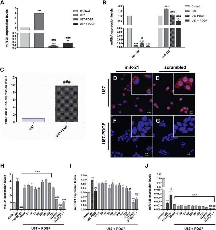
Figure 1.
PDGF-B-related modulation of miRNA-expression levels in U87 and U87-PDGF human GBM cells. (A) MiR-21 and (B) miR-128/miR-221 expression levels in parental U87 cells, PDGF-B-overexpressing U87 cells (U87-PDGF) and U87 cells cultured in the presence of 30 ng/ml PDGF-B for 60 days (U87 + PDGF), compared with control epileptic tissue. (C) PDGF-B mRNA levels in U87 and U87-PDGF. FISH staining in cultured (D and E) U87 and (F and G) U87-PDGF cells. Cells were stained with (D and F) miR-21 or (E and G) noncoding (scrambled) probes. Nuclear staining was accomplished using the cell-permeable DNA stain Hoescht 33342 (Blue). MiR-21 staining (red dots) is observed in U87 cells, predominantly in the cytoplasm, whereas only residual staining is detected in U87-PDGF cells. Control experiments targeting the endogenous U6snRNA (positive control) or without LNA probe (negative control) were performed in parallel (Supplementary Material, Figure S2D–G). Images were obtained by confocal microscopy with a 40× EC Plan-Neofluar. Scale corresponds to 10 µm. (H) MiR-21, (I) miR-221 and (J) miR-128 expression levels after culturing parental U87 cells in medium supplemented with PDGF-B (30 ng/ml) for different time periods; 21 days + 1: cells cultured for 21 days in PDGF-B-enriched medium followed by 7 days of culture in normal medium (PDGF-B-depleted medium). *P < 0.05, **P < 0.01, ***P < 0.001 compared with control epileptic tissue. #P < 0.05, ##P < 0.01, ###P < 0.001 compared with parental U87 cells.

