Figure 2.
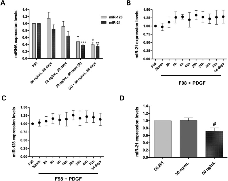
PDGF-B-related modulation of miRNA-expression levels in F98 rat and GL261 mouse glioma cells. (A) MiR-21 and miR-128 expression levels in F98 cells cultured in PDGF-B-depleted medium (control) or cultured in medium supplemented with PDGF-B (30 or 50 ng/ml) for different time periods; (A) +50 ng/ml, 30 days: cells cultured for 60 days in PDGF-B-enriched medium (30 ng/ml), followed by a further incubation for 30 days in medium containing a higher concentration of PDGF-B (50 ng/ml). (B) MiR-21 and (C) miR-128-expression levels in F98 cells cultured in medium supplemented with PDGF-B (30 ng/ml) for different time periods, compared with those obtained in F98 cells cultured in PDGF-B-depleted medium. 14 days: cells cultured for 14 days in PDGF-B-enriched medium. (D) MiR-21-expression levels in GL261 glioma cells cultured in PDGF-B-depleted medium (control) or cultured in medium supplemented with PDGF-B (30 or 50 ng/ml) for 30 days. *P < 0.05, **P < 0.01, ***P < 0.001 compared with control F98 cells; #P < 0.05 compared with control GL261 cells.

