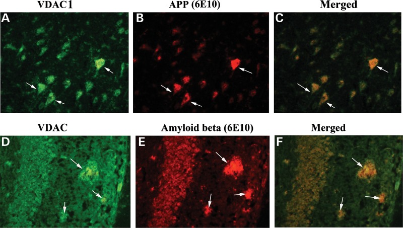
Figure 8.
Double-labeling immunofluorescence analysis of VDAC1, Aβ and full-length APP in cortical sections from 20-month-old APP mouse. The localization of VDAC1 (A) and APP (B) and the colocalization of VDAC1 and APP (merged C) at 40× original magnification; the localization of VDAC1 (D); Aβ deposits (E); and the colocalization of VDAC1 and Aβ deposits (merged F) at 100× original magnification, in hippocampal sections from 20-month-old APP mice.

