Figure 1. .
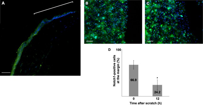
The expression of Notch1 was evaluated in the mouse corneal epithelium at 6 hours after a debridement wound. Notch1 staining was distinctly reduced in the cells near the leading edge (the bracket) (A). The expression of Notch1 was evaluated in primary human corneal epithelial cells subjected to a scratch assay at baseline (B) and after 12 hours (C). The percentage of cells that stained for Notch1 in the leading edge was 42% lower at 12 hours compared with immediately (0 hour) after the scratch (D) (P < 0.001). (Green: FITC, blue: DAPI); scale bar is 40 μm for (A) and 100 μm for (B) and (C). Asterisk represents statistically significant data.

