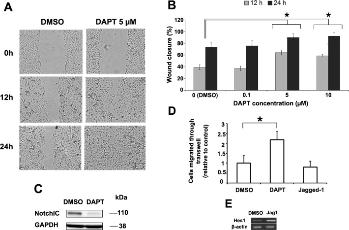
Figure 2. .
Human corneal epithelial cells were subjected to a scratch assay and then treated with DAPT or DMSO (control) (A). The effect of DAPT concentration on scratch assay wound closure rate was measured (B). * Represents a statistically significant difference compared to control (P < 0.001). Western blot for Notch1IC confirmed that 10-μM DAPT can effectively inhibit Notch activation (C). HCE-T cells pretreated with DAPT migrated 2.2 times faster than control in transwell migration assay (P < 0.0001) while Jagged1 treated cells migrated 20% slower but did not reach statistical significance (P = 0.077) (D). Jagged1 treatment was found to activate Notch by inducing the expression of its downstream Hes1 (E). Asterisk represents statistically significant data.

