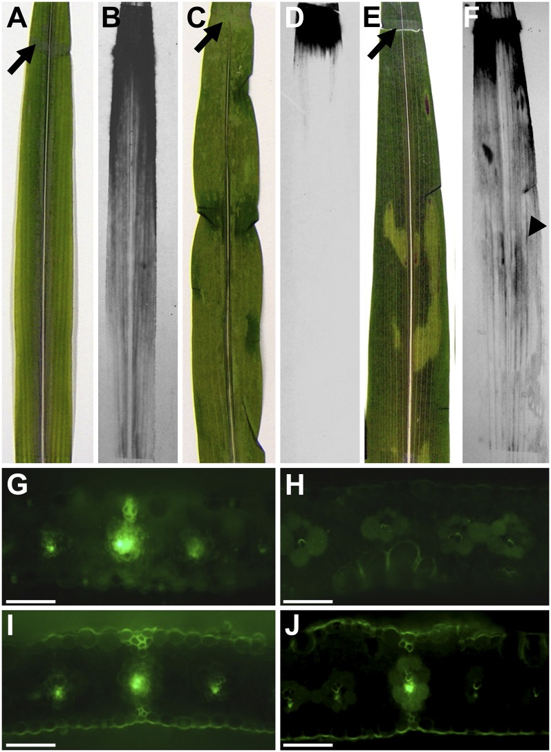
Figure 4.
14C-labeled Suc and CF transport in wild-type and tdy2 mutant leaves. A, C, and E, Black arrows indicate sites of abrasion and application of [14C]Suc to leaves. B, D, and F, Autoradiographs showing [14C]Suc localization in leaves 1 h after application. A and B, Wild-type leaf showing normal phloem loading and transport of [14C]Suc. C and D, A mostly yellow tdy2 mutant leaf in which 14C-labeled Suc was applied to a yellow region, showing that labeled Suc did not move from the site of application. E and F, tdy2 mutant leaf in which labeled Suc was applied to green tissue distal to the yellow tissue, showing that labeled Suc was loaded, translocated long distance in the phloem, and passed through the phloem in the yellow tissue (arrowhead). G to J, CF transport assays in which CFDA was applied to the leaf tissue near the tip; cross-sections were taken 5 to 7 cm proximal to the application site. Bright green fluorescence shows the presence of CF in the phloem. G, Wild-type leaf showing normal phloem translocation. H, A mostly yellow tdy2 mutant leaf in which CFDA was applied to distal yellow tissue; a cross section was taken from a proximal yellow region, showing that CF was not able to be translocated through the phloem. I, tdy2 mutant in which CFDA was applied to distal green tissue; a cross-section was taken from proximal green tissue, showing normal phloem translocation. J, tdy2 mutant tissue in which CFDA was applied to distal green tissue; a cross-section was taken from proximal yellow tissue, which shows that CF can enter the phloem translocation stream in the green tissue and subsequently be translocated through yellow tissues, indicating that long-distance phloem transport is unobstructed. Bars + 100 µm. [See online article for color version of this figure.]

