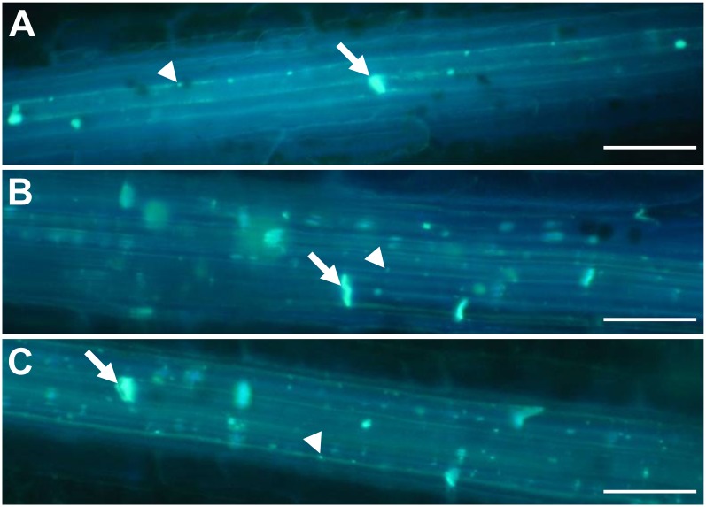
Figure 7.
Aniline blue staining of callose in wounded leaves. A, The wild type. B, tdy2-R green leaf region. C, tdy2-R yellow leaf region. Images show callose staining using aniline blue 10 min after removal of the epidermal surface. The callose is visualized as bright blue fluorescence. White arrows identify callose at the sieve plates, and white arrowheads identify callose deposition at PD along the SE cell wall. Bars + 50 μm. [See online article for color version of this figure.]

