Figure 2.
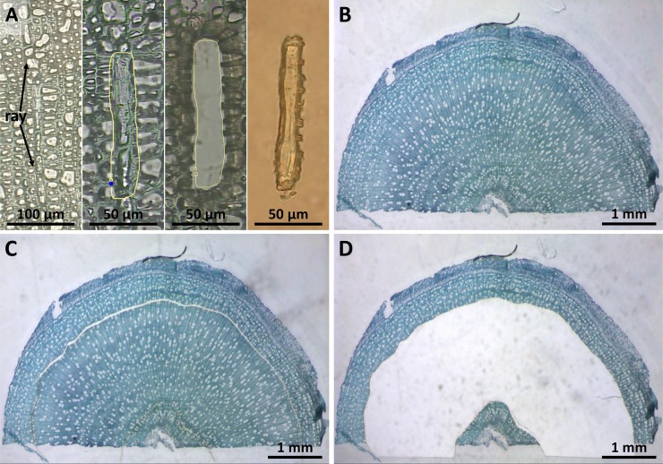
LMPC of ray cells from poplar wood. A, First LMPC attempt of rays. From left to right: selected ray, signed ray, remains after ray dissection, catapulted ray. RNA yield and quality from these samples were not adequate for microarray hybridization. B to D, Inverse LMPC. B, Cross section of a poplar twig. C, Dissection of the area between the xylem differentiation zone and the central section. D, Remains after removal of ray-enriched wood. [See online article for color version of this figure.]

