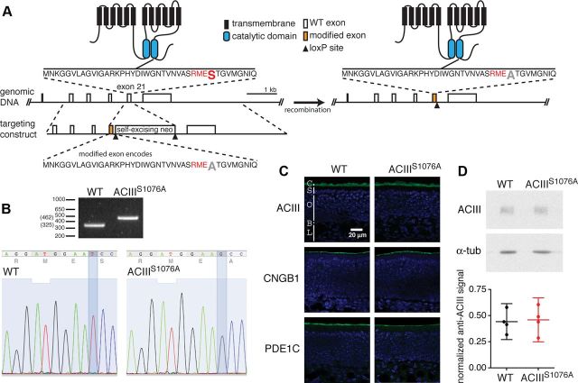
Figure 1.
Generation of ACIIIS1076A mice. A, Illustration of the genetic targeting strategy. Left, Wild-type ACIII protein structure (top), partial Adcy3 gene structure with open boxes representing exons (middle), and the targeting construct (bottom). The orange boxes in the targeting construct and in the genomic DNA (right) represents the modified exon 21 containing codon mutation from serine1076 to alanine. The amino acid sequence coded by exon 21 with the CaMKII consensus sequence RMES in red and the serine1076 in a larger font, is shown and its location in ACIII protein is indicated. Right, The corresponding modified ACIII protein and gene structure. B, Top, PCR-based genotyping of wild-type and ACIIIS1076A homozygous littermates. Bottom, Sequencing confirms the T-to-G base pair substitution in ACIIIS1076A mice, changing the codon of serine (TCC) to that of alanine (GCC). C, Immunostaining on sections of OE for ACIII, CNGB1b, and PDE1C. Sections were counterstained with DAPI (blue) to label cell nuclei. C, Cilial layer; S, supporting cell layer; O, OSN layer; B, basal lamina; L, lamina propria. D, Top, Western blots for ACIII and α-tubulin (α-tub) on total OE proteins. Bottom, Quantification of Western blotting. Values are normalized to α-tubulin staining and shown in arbitrary units. ACIII is detected at similar levels in wild-type and ACIIIS1076A mice. Each datum point represents an individual mouse. Error bars are 95% CI.

