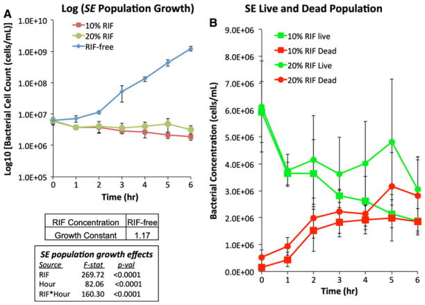
Fig. 4.
Time-course measurements of suspended cell concentrations of Staphylococcus epidermidis cultures (5.4 × 10⁷ cells/mL) exposed to various scaffolds for 6 h at 37°C. a Total suspended cell concentrations exposed to RIF-free (diamond), 10%-RIF (square) and 20%-RIF (circle) scaffolds. (b) Live (green) and dead (red) suspended cell concentrations for cultures exposed to 10%-RIF (square) and 20%-RIF (circle) scaffolds

