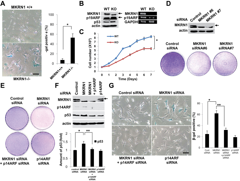
Figure 2.
Knockout or knockdown of MKRN1 induces senescence in MKRN1 KO MEF and HFF cells through the stabilization of ARF. A) Premature senescence was induced in MKRN1−/− MEF. MKRN1−/− and +/+ MEFs were stained for SA-β-galactosidase activities (×200 magnification). The graph shows the percentage of β-galactosidase-positive cells. The error bars indicate 95% confidence intervals. *, P = .005 using two-sided t test. Three independent experiments were performed. The scale bar indicates 160.51 µm. B) p19ARF was stabilized in MKRN1−/− MEFs. Cell lysates of MKRN1−/− and +/+ MEFs were detected with anti-p19ARF, p53, and MKRN1 antibodies. Cells were analyzed by RT-PCR using primers specific for p19ARF, MKRN1, and GAPDH. Arrows indicate MKRN1 and p19ARF proteins. C) MKRN1−/− MEFs showed growth retardation. The same amount of MKRN1 +/+ and −/− MEFs was plated and counted 1, 2, 3, 4, 5, 6, and 7 days later. The error bars indicate 95% confidence intervals. *, P = .003 using two-sided t test. Three independent experiments were performed. D) Knockdown of MKRN1 suppresses growth of HFF cells. After 96-h transfection with control, MKRN1 #6, or MKRN1 #7 siRNAs, the cells were stained with crystal violet. Cell lysates were subjected to immunoblotting using anti-MKRN1 and actin antibodies. The arrow indicates MKRN1. E) p14ARF depletion blocks the inhibition of cell growth induced by MKRN1 ablation. HFF cells were transfected with control or MKRN1 #6 siRNA, with or without p14ARF siRNA. After 96h, cells were stained with crystal violet. F) p14ARF protein is stabilized by MKRN1 depletion. HFF cells were treated as indicated, and their lysates were subjected to western blotting using antibodies against p14ARF, MKRN1, p53, and actin. The arrow indicates MKRN1. The p53 protein level was quantified using Image-J software. The error bars indicate 95% confidence intervals. *, P = .01; **, P = .03 using two-sided t test. Three independent experiments were performed. G) Cellular senescence induced by MKRN1 depletion is reversed by p14ARF ablation. HFF cells were transfected with control or MKRN1 #6 siRNA, with or without p14ARF siRNA, as indicated. After 96h, cells were stained for β-galactosidase activities (×200 magnification). The scale bar indicates 160.51 µm. The graph shows percentage of β-galactosidase-positive cells. The error bars indicate 95% confidence intervals. *, P = .008; **, P = .002 using two-sided t test. Three independent experiments were performed. MKRN1 = Makorin ring finger protein 1; p14ARF = p14 alternative reading frame; p19ARF = p19 alternative reading frame; MEFs = mouse embryonic fibroblasts; HFF = human foreskin fibroblast.

