Figure 3.
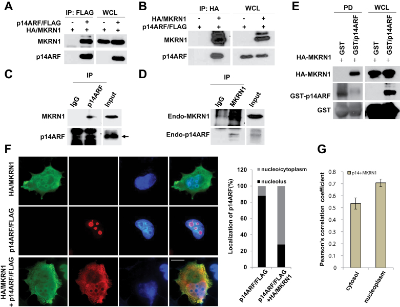
MKRN1 interacts with p14ARF. A, B) MKRN1 and p14ARF proteins interact with each other. Plasmids expressing HA/MKRN1 alone or with p14ARF/FLAG were transfected into 293 cells. WCL were immunoprecipitated with anti-FLAG or anti-HA antibodies, and immunoprecipitates and WCLs were immunoblotted using the antibodies against MKRN1 and p14ARF. C, D) Endogenous MKRN1 and p14ARF bind to each other. HeLa cell lysates were immunoprecipitated with anti-p14ARF or MKRN1 antibodies, and immunoprecipitated proteins were analyzed using the same antibodies. Arrow indicates p14ARF. E) Recombinant p14ARF and MKRN1 proteins bind to each other directly. Purified GST/p14ARF and in vitro translated HA-MKRN1 were incubated at 37°C (input) followed by pull-down (PD) using glutathione-sepharose. The precipitated samples were detected using anti-MKRN1 and p14ARF antibodies. F) MKRN1 induces p14ARF translocation to nucleo/cytoplasm. H1299 cells were transfected with plasmids expressing HA/MKRN1 and p14ARF/FLAG. Fixed cells were detected by monoclonal anti-HA mouse and polyclonal anti-FLAG rabbit antibodies and stained by Alexa Fluor 594-conjugated anti-rabbit and Alexa Fluor 488-conjugated anti-mouse antibodies, analyzed using N-SIM superresolution microscopy (×150 magnification). The percentages on panels 6 and 10 and graph indicate the fractions of counted cells (200) that displayed such p14ARF/FLAG localization. G) The Pearson correlation coefficient for the colocalization of p14ARF and MKRN1. The error bars indicate 95% confidence intervals using two-sided t test. Thirty cells were tested for the analyses. The tan bars show the Pearson correlation for p14ARF and MKRN1 colocalization. MKRN1 = Makorin ring finger protein 1; p14ARF = p14 alternative reading frame.

