Figure 7.
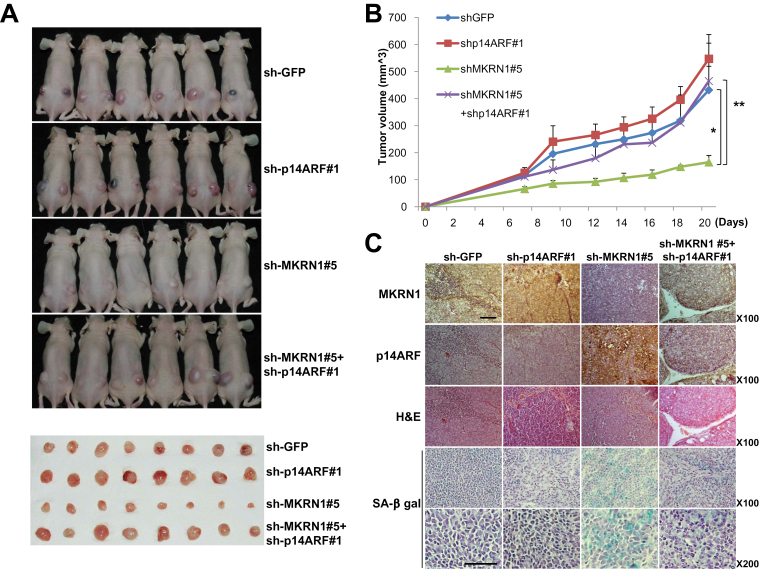
The suppressive effect of MKRN1 on tumor growth was rescued by p14ARF depletion. A–C) Mice (N = 6) were inoculated subcutaneously into both flanks with 106 cells of each AGS cell line. Lentiviruses expressing sh-RNA targeting p14ARF, MKRN1, or both were employed to produce stable AGS cell lines. Lentivirus expressing sh-RNA targeting GFP was used as a control. A) At 23 days after inoculation, mice were killed in 7.5% CO2 chamber, and mice and excised tumors from mice were photographed. B) Quantification of tumor formation was performed by measurement of tumor size 7, 9, 12, 14, 16, 18, and 20 days after inoculation (N = 6). The error bars indicate 95% confidence intervals. *, P = .001; **, P = .009, two-sided t test. All statistical tests were two-sided. C) Immunohistochemical analysis and hematoxylin and eosin (H&E) staining were performed for each group of tumors as described in “Materials and Methods” (magnification ×100). SA-β-galactosidase-stained slides were viewed under ×100 and ×200 magnification. The scale bar indicates 100 µm. MKRN1 = Makorin ring finger protein 1; p14ARF = p14 alternative reading frame.

