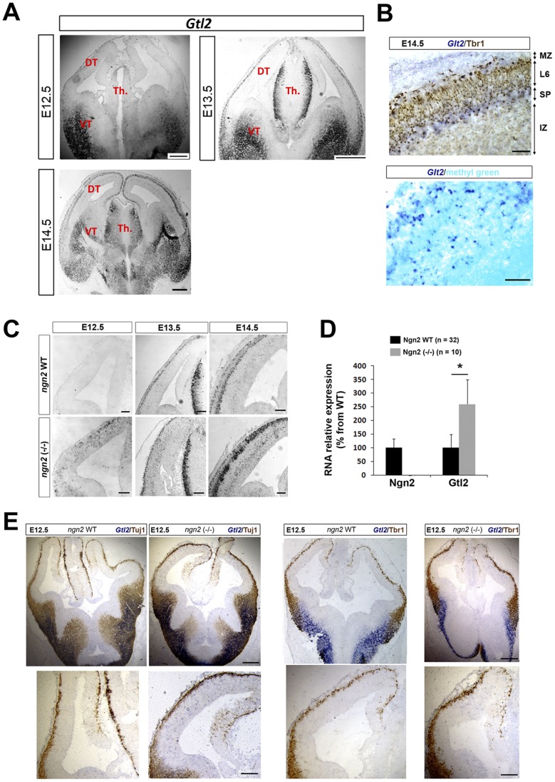
Figure 1. Gtl2 is upregulated and ectopically expressed in the dorsal telencephalon of Ngn2 KO mice. A.
Detection of Gtl2 RNA at E12.5, E13.5 and E14.5 by ISH in WT mice. DT = dorsal telencephalon, VT = ventral telencephalon, Th. = thalamus. B. Expression of Gtl2-positive neurons in the subplate (SP) and marginal zone (MZ) but not in Tbr1 (layer 6) positive neurons of the developing cortical plate (upper panel). Gtl2 ISH counterstained with the nuclear marker methyl green in the cortex (lower panel). C. Gtl2 expression (ISH) in the dorsal telencephalon of WT and Ngn2 KO mice at E12.5, E13.5, and E14.5. D. Overexpression of Gtl2 in Ngn2 KO dorsal telencephalon at E13.5 assessed by qPCR. Data are presented as percentage of change normalized to the mean of WT (taken as 100%) + s.e.m; *P<0.01 in Mann-Whitney’s test. E. ISH of Gtl2 co-labeled with Tuj1 (4 left panels) and Tbr1 (4 right panels) proteins (immunohistochemistry) shows that Gtl2 is ectopically expressed in Tuj1 and Tbr1 negative regions in Ngn2 KO mice. Scale bars: A: 400 µm. B: 50 µm. C and E upper panels: 200 µm, C and E lower panels: 50 µm.

