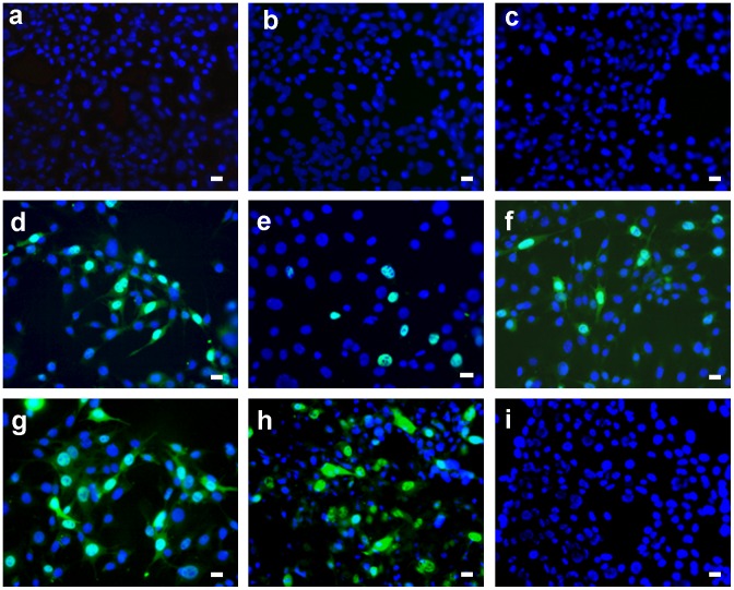
Figure 5. Inactivation of the mouse polyomavirus on the surface of TPP-doped Tecophilic® nanofiber textile.
Cells infected with polyomavirus eluate from the surface of the nanofiber textile after 30 minutes of irradiation (a, b, c) or without irradiation (d). Cells infected with control polyomavirus eluate from the surface of the textile without TPP after 30 minutes of irradiation (e) or without irradiation (f). Cells infected with the same amount of the virus in the absence of the textile after 30 minutes of irradiation (g) or without irradiation (h). Non-infected cells (i). Detection of the LT antigen (green) in the nuclei of infected cells. To visualize cell nuclei, DNA was stained with DAPI (blue). Representative images are shown with the bar of 20 µm at the right corner.

