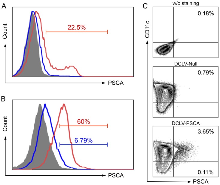
Figure 1. Targeted transduction and delivery of PSCA antigen gene into dendritic cells (DCs) by DCLV-PSCA.
(A) 293T cells were transfected transiently with plasmids FUW-Null (mock control, blue line) or FUW-PSCA (red line). Two days later, cells were collected and stained for PSCA expression analyzed by flow cytometry. 293T cells stained with the isotype antibody were included as a control (grey shade area). (B) 293T cells were transfected transiently with plasmids FUW-PSCA, SVGmu, and other necessary lentiviral packaging plasmids to produce DCLV-PSCA vectors. Fresh virus supernatant was used to transduce 293T cells (blue line) or 293T.hDC-SIGN cells (red line) with MOI = 10. PSCA expression was analyzed by flow cytometry 3 days post-transduction. (C) Bone marrow-derived DCs were transduced with a mock vector DC-LV-Null or DC-LV-PSCA vector. Five days later, CD11c and PSCA expression were assessed by flow cytometric analysis. All experiments were repeated three times and the representative data is shown.

