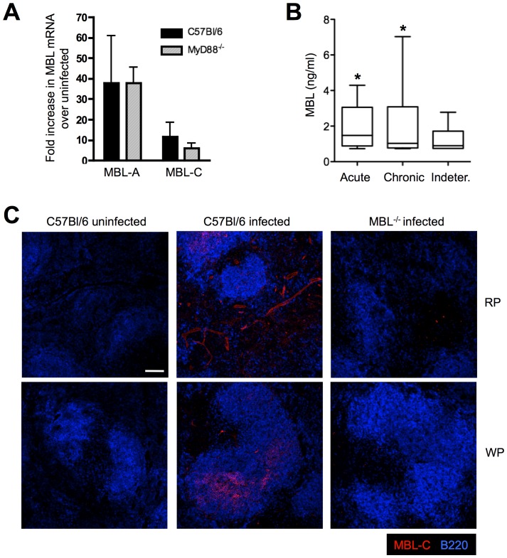
Figure 1. T. cruzi infection induces MBL expression in vivo.
WT and MyD88−/− mice were infected with trypomastigotes of the Y strain of T. cruzi and 9 days after infection, MBL-A and MBL-C mRNA accumulation was determined in spleens from infected and uninfected animals by real-time PCR (A). Detection of MBL-C protein was also performed by immunofluorescence microscopy in splenic sections obtained from WT mice infected as above (B). Micrograph shows MBL-C (red) and B220 (blue) staining in both the red (RP) and white pulp (WP). Scale bar, 100 µm. Infected MBL−/− spleens were included as a control. All panels shown are representative of 2 independent experiments using 3 to 4 mice per group. Bars indicate the standard error of the mean (SEM). Sera from patients with acute, chronic or indeterminate Chagas’ disease were assayed for MBL as described in Material and Methods (C). *, Indicates statistically significant differences between acute vs indeterminate Chagas’ disease groups.

