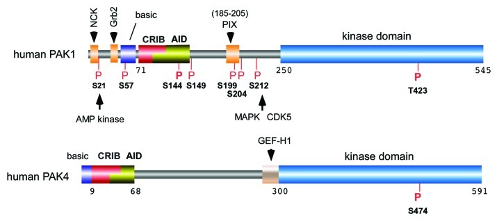
Figure 1. The domain structure of PAK1 and PAK4 highlighting the conserved features of group I kinases and the conserved sites of kinase phosphorylation. The presence of proline-rich SH3 binding sites are marked in orange. The p21-binding domain (PBD or CRIB) is indicated in red and overlaps the auto-inhibitory domain (AID) in yellow. The basic residue cluster required for phospholipid-mediated kinase activation is marked in purple. PAK1 phosphorylation sites that are conserved across other isoforms are marked in red. The activation-loop phopho-residue indicated: this is constitutively phosphorylated in the case of PAK4. Unless otherwise indicated these are auto-phosphorylation sites.

An official website of the United States government
Here's how you know
Official websites use .gov
A
.gov website belongs to an official
government organization in the United States.
Secure .gov websites use HTTPS
A lock (
) or https:// means you've safely
connected to the .gov website. Share sensitive
information only on official, secure websites.
