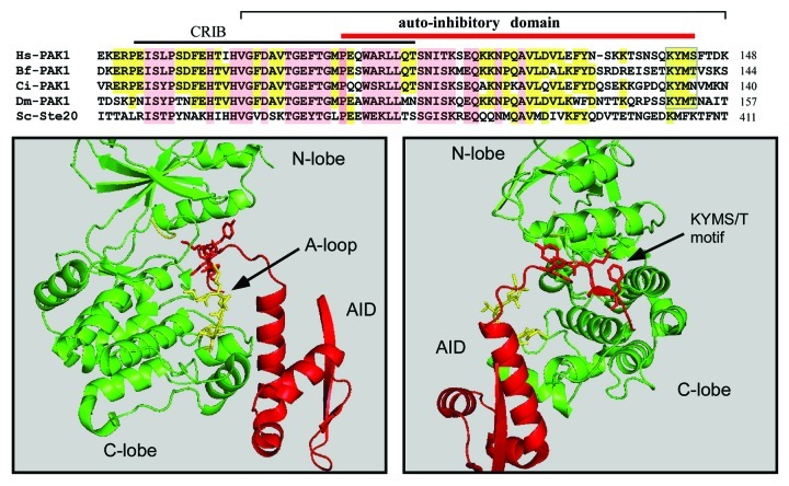
Figure 2. The structure of the PAK1 AID and sequence alignment among Group I PAKs from diverse phyla. PAK1 sequences are from human (Hs, Q13153), Branchiostoma floridae (Bf, XP_002595185), Ciona intestinalis (Ci, XP_002131099), dPAK1 from Drosophila melangaster (Dm, AAC47094) and Ste20p from Saccharomyces cerevisiae (Sc, AAA35038). Completely conserved residues are in pink and partial conservation in yellow. The interaction of the KYMS box is illustrated in the figure below, which shows a complex between human PAK1 and the AID (PDB: 1F3M). The A-loop in yellow is displaced by the presence of the KYMS sequence, which occupies a position under the α-C helix. The structure was prepared using Pymol.

An official website of the United States government
Here's how you know
Official websites use .gov
A
.gov website belongs to an official
government organization in the United States.
Secure .gov websites use HTTPS
A lock (
) or https:// means you've safely
connected to the .gov website. Share sensitive
information only on official, secure websites.
