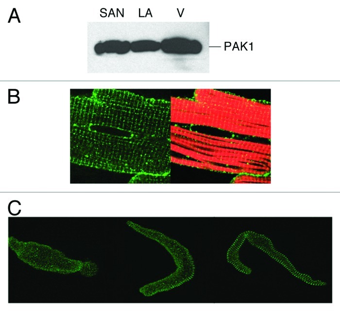
Figure 1. PAK1 expression in the heart. (A) Western blot detection of PAK1 expression in SAN, left atrial (LA) and ventricular tissues (V). PAK1 expression pattern was examined by immunocytochemical staining in (B) adult rat cardiomyocyte (green, anti-PAK1; red, rhodamine-conjugated phalloidin), in (C) guinea pig senatorial node (SAN) cells (adapted from refs. 3 and 4).
