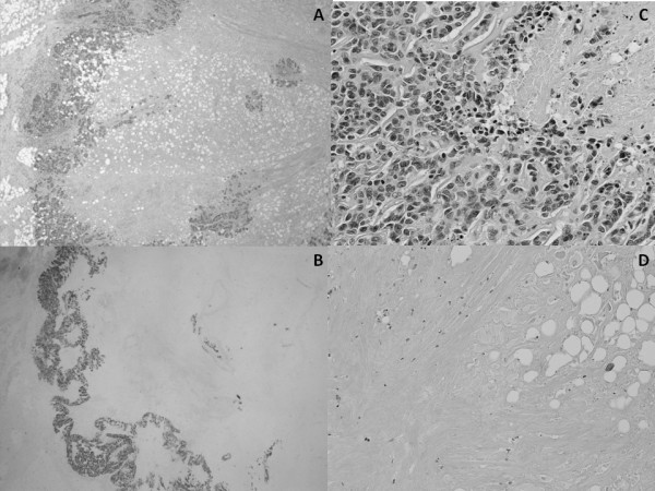
Figure 4.

Microscopic images from pathological study.A. The tumor shows a unicentric nodule with a prominent, extensive central hipocellular zone, surrounded by a narrow ring of viable tumor cells. B. E-cadherin positivity of the malignant cells strongly shows the narrow rim of viable tumor. C. Peripheral rim. The tumor cells display high nuclear grade with numerous mitoses. D. Central, hypocellular zone with fat necrosis and formation of fibrotic and hyaline material.
