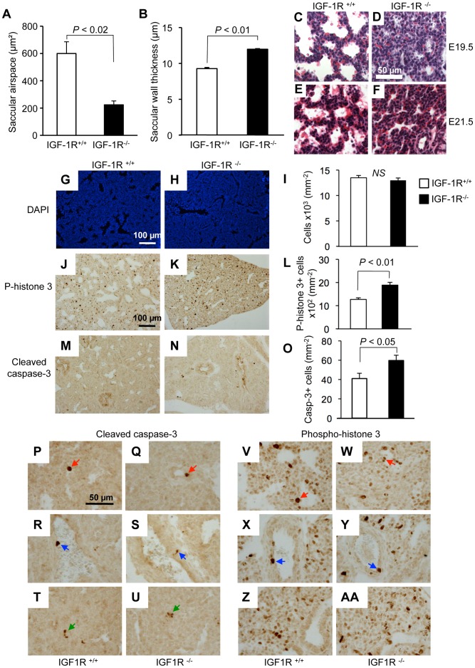
Figure 4. Lung histomorphology and cell turnover in the absence of IGF-1R.
A, Saccular airspace and B, saccular wall thickness (mean ± SEM) at developmental stages E17.5 in IGF-1R+/+ (n = 4) and IGF-1R−/− embryos (n = 4). Wilcoxon Mann-Whitney U test. C–F, Extended gestation period and lung histology. H&E stain of lung tissue from embryos at 19.5 (C, D) and 21.5 days (E, F). To extend gestation period up to 21.5 days, pregnant mothers were treated with progesterone from E17.5 onwards. Note the presence of red blood cell extravasation in E21.5 lung samples from IGF-1R+/+ and IGF-1R−/− mice. G–O, Cell turnover in IGF-1R−/− embryonic lung at E17.5. Lung histology from IGF-1R+/+ embryos (G, J and M) and IGF-1R−/− embryos (H, K and N) at E17.5. Bar graphs (I, L and O) show quantification (mean ± SEM; n = 3–7 individuals per group; Student’s t-test). G–I, Cells were counted using DAPI staining (blue signal). J–L, Cell proliferation was measured using phospho–histone H3 immunohistochemistry (brown staining). M–O, Apoptosis was detected using cleaved caspase-3 immunohistochemistry (brown staining). Cleaved caspase-3 (P-U) and phospho-histone H3 labeling (V-AA) at high magnification showing examples for IHC-positive epithelial (red arrows), vascular endothelial (blue) and mesenchymal cells (green), as identified by their anatomical location. Note that many of the proliferating cells are located in areas that are composed of mostly mesenchymal cells.

